Figure 5.

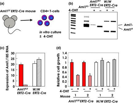
Depletion of AML1 inhibits the growth of CD4 positive T‐cells. (a) Splenic T‐cells of Aml1 fl/flERT2‐Cre mice were isolated using an antibody against CD4 and cultured for 5 days with or without 4‐hydroxytamoxifen (4‐OHT) in the presence of IL‐2. (b) Genotype of Aml1 was analyzed after 5‐day culture with or without 4‐OHT in the presence of IL‐2. Primers used for genotyping were as follows: f2: ACAAAACCTAGGTGTACCAGGAGAACAAGT, r1: GTCTACTCCTTGCCTCAGAAAACAAAAAC, fl20: CCCTGAAGACAGGAGAAGTTTCCA. (c) Expression of Aml1 was measured by quantitative reverse transcription‐polymerase chain reaction (qRT‐PCR) and correlated to 18S rRNA levels after 5‐day culture with or without 4‐OHT in the presence of IL‐2. (d) T‐cells with or without the Aml1 allele were cultured in the presence of IL‐2 for 10 days. The growth rate (live cells) was measured by the production of adenosine triphosphate (ATP). ATP production of cells from Aml1fl/flERT2‐Cre mice (red bars, diagonal stripe) treated with 4‐OHT was less than that of untreated cells (red bars, filled). The drug was not toxic to wild‐type cells (white and gray diagonal stripes: treated and non‐treated, respectively).
