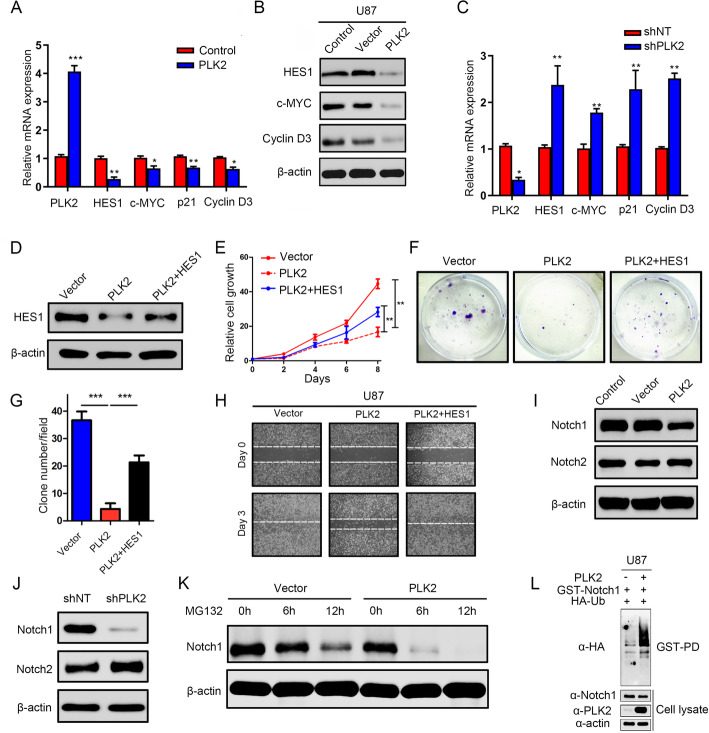
Fig. 6.
Loss of PLK2 enhances TMZ resistance of GBM via activation of Notch signaling. a, qRT-PCR assays were performed to measure the mRNA expression levels of the downstream targets of Notch signaling when cells were pre-transduced with lentiviral PLK2 and its negative control. b, Western blot analyses were conducted to detect the protein levels of downstream targets of Notch signaling. β-actin was used as an internal control. c, qRT-PCR assays were carried out to measure the mRNA expression levels of Notch downstream targets when treated with lentiviral shPLK2. d, Western blot assays was used to detect the protein level of HES1 in PLK2 OE U87 cells when transduced with HES1 lentivirus. e, Time survival curve of U87 cell pretreated with indicating interventions. f and g, Colony formation ability of U87 cells pretreated with different lentiviruses as indicated ((***P < .001, with student’s t-test). h, representative images of wound healing assays to indicate the migratory ability of U87 cells pretreated with different lentiviruses. i, the effect of PLK2 OE on activated Notch1 and Notch2 protein was detect by western blot, β-actin was used as an internal control. j, the effect of PLK2 knockdown on activated Notch1 and Notch2 protein was detected by western blot, β-actin was used as an internal control. k, the effect of proteasome inhibitor MG132 on protein level of Notch1 pretreated with or without PLK2 OE lentivirus, β-actin was used as an internal control. l, GST-pulldown assay showed that the ubiquitination level of GST-Notch1 increased when PLK2 was overexpressed compare with control group in U87 cell line. All data were presented as the mean ± SD of triplicate independent experiments

