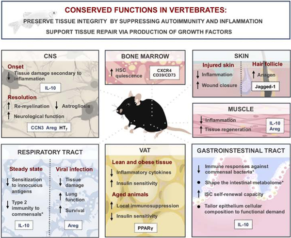
Figure 1: Roles of regulatory T (Treg) cells in tissue pathophysiology and metabolism in vertebrates.

Conserved functions of Treg cells across vertebrate species (top blue box). Specific functions of Treg cells in various tissues are summarized in bottom panels with molecules implicated in corresponding Treg cell functions highlighted in white boxes. Secreted molecules depicted in blue; surface or intracellular molecules depicted in black. Asterisks (*) denote functions ascribed to extrathymically generated Treg cells. From top left to right: Treg cells in the central nervous system (CNS) limit secondary tissue damage and contribute to tissue repair during ischemia and spinal cord injury. Treg cell production of adenosine via ectonucleotidases CD39 and CD73 has been suggested to support the quiescence of hematopoietic stem cells (HSC) in the bone marrow. Skin Treg cells expressing a Notch receptor ligand Jagged-1 promote wound closure through a contact-dependent Notch signaling in stem cells to promote anagen. Treg cells infiltrate the muscle upon injury and suppress inflammation, facilitating tissue repair and regeneration. Intestinal Treg cells shape the composition and function of the microbiota and affect the make-up of the gut epithelium via direct and indirect effects on intestinal stem cells (ISC). Dual role of Treg cells in visceral adipose tissue (VAT) function: in young animals, Treg cells promote insulin sensitivity in both lean and obese tissue, while a buildup of fat Treg cells in older animals contributes to insulin resistance. In the lung, Treg cells support tissue homeostasis by preventing exacerbated immune responses to innocuous environmental antigens at the steady state and by partaking in tissue repair during injury.
