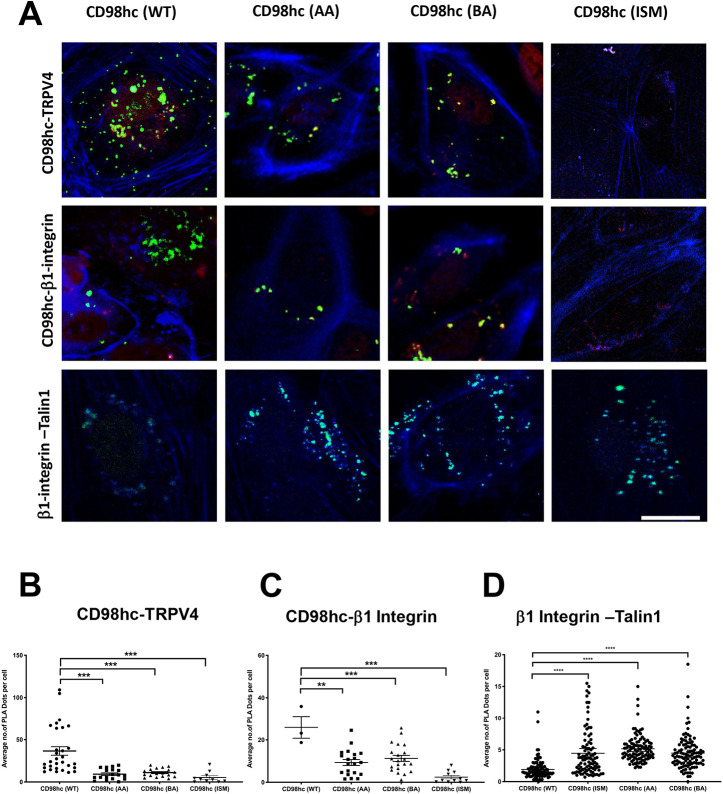
Fig. 5.
Proximity profiling of β1-CD98hc-TRPV4 interactions. (A) Confocal micrographs of basal slices of HUVE cells showing proximity ligation foci (green) between FLAG-CD98hc wild type (WT), mutated acidic residues (AA) or mutated basic residues (BA) and endogenous TRPV4 (top row), endogenous β1 integrin (middle row) or endogenous β1 integrin and endogenous Talin1 (bottom row). Phalloidin staining of F-actin is shown in blue. Scale bar: 20 μm. (B) Scatter plots showing individual number of PLA dots per cell within basal sections of images from five fields of view per biological replicate, corresponding to experiments shown in A (n=3; **P<0.01, ***P<0.001, ****P<0.0001; bars indicate the mean with 95% CI).

