FIG 5.
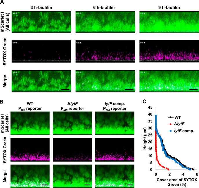
Dead cells and eDNA are also abundant near the bottom of the biofilm. (A) The Pldh reporter was grown in a glass-bottom dish in BHIs with sCSP at 37°C. The medium was supplemented with 1.25 μM SYTOX Green to stain dead cells and extracellular nucleic acids. The same field of view within a range of approximately 40 μm from the glass surface was observed while culturing at 37°C. Z-stacks were acquired at 0.55 μm intervals. These images show side views of the biofilms, and the white line below each image shows the glass surface obtained by the reflection. (B) The WT, lytF deficient mutant (ΔlytF), and lytF-complemented (lytF comp) strains with a Pldh reporter background were grown in an aerobic atmosphere containing 5% CO2 at 37°C in BHIs with sCSP for 6 h in glass-bottom dishes. Planktonic cells were removed by washing twice with PBS. These images show side views of the biofilms. Z-stacks were acquired at 0.5 μm intervals. mScarlet-I (all cells) and SYTOX Green (dead cells and extracellular nucleic acids) are shown in green and magenta, respectively. The scale bars indicate 10 μm. Representative images from independent experiments are presented. (C) Image analysis of the 3D images shown in panel B. In each two-dimensional image, the cover areas showing the fluorescence of SYTOX Green were calculated by ImageJ and Fiji.

