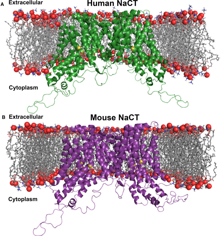
Figure 8. Homology modeling of human and mouse NaCT.
(A) Ribbon representation of the human NaCT homodimer in the inward-facing conformation. The homology model was embedded in a DOPC phospholipid bilayer for molecular dynamics simulation. (B) Ribbon diagram of the mouse NaCT homodimer in the inward-facing conformation embedded in a DOPC phospholipid bilayer. Red spheres are phosphates and grey sticks are the diacylglycerol backbone of phosphatidylcholine, the most abundant lipid in animal cell membranes. Each monomer forms an internal cavity with a Na+ (yellow spheres) and citrate (cyan sticks), adjacently bound to each pocket at the cytosolic basin of the protomer. Citrate ions are exposed to the cytosolic space whereas the Na+ ions are buried.

