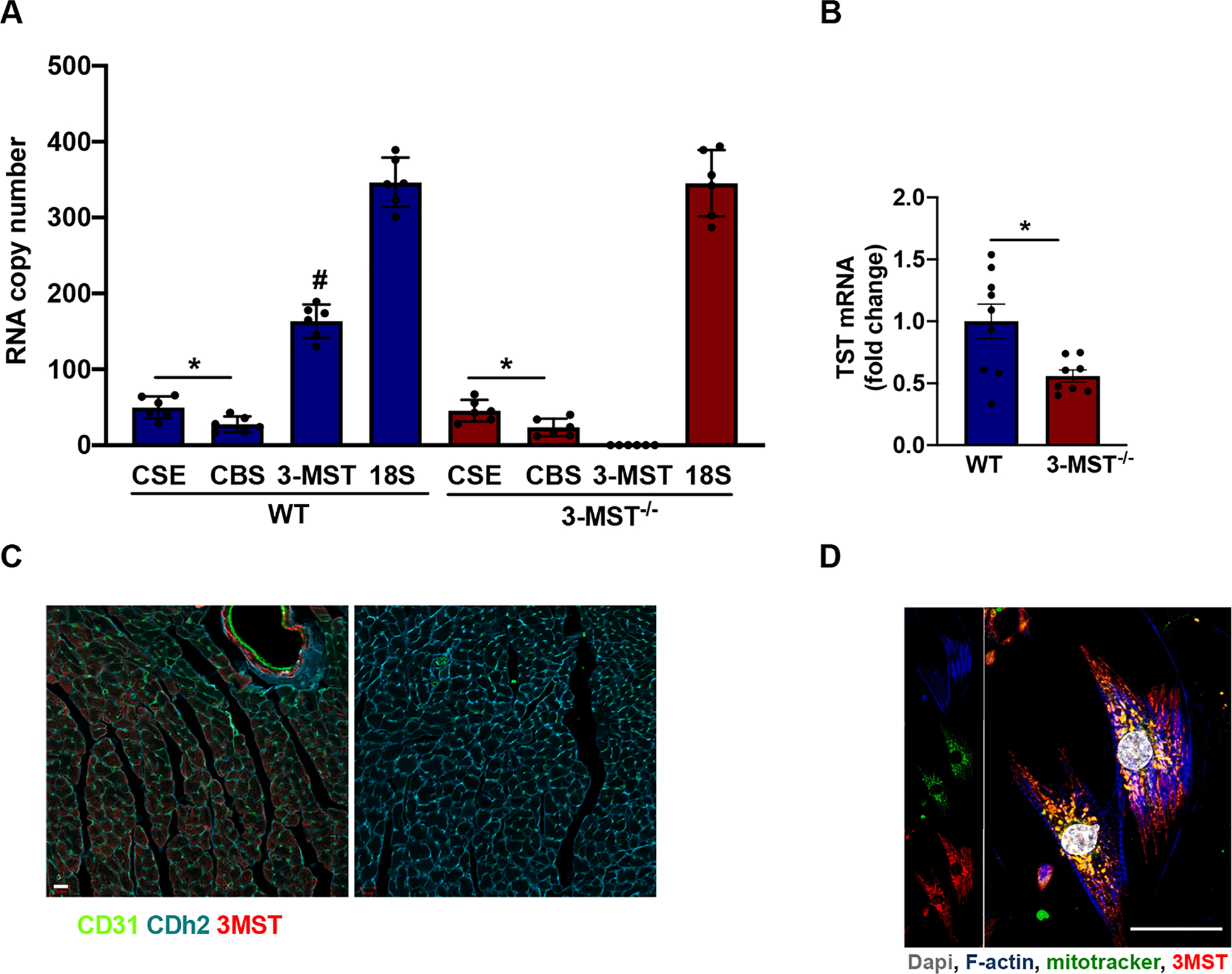
Fig. 1.

Expression of 3-MST in the heart. mRNA copy number of H2S producing enzymes (CSE, CBS, 3-MST) compared to the copy numbers of 18S rRNA in the heart of WT and 3-MST−/− mice (A). Steady state mRNA levels of TST in the heart of WT and 3-MST−/− mice (B). Immunostaining of CD31 (green, endothelial cell marker), Cdh2 (blue, cardiomyocyte marker) and 3-MST (red) in WT heart cross-sections. Comparable results were obtained in 5 additional animals, bar = 20 μm (C). Subcellular localization of 3-MST in cardiomyocytes. Representative immunostaining of primary isolated murine cardiomyocytes from WT hearts. F-actin (blue), Dapi (grey), mitotracker (green) and 3-MST (red). The co-localization of 3-MST and mitotracker signals was approximately 60% of the total, bar = 25 μm. The panel on the right is a negative control (no primary Ab) for the 3-MST staining (D). Values are shown as mean ± SEM, n = 6/group (A), n = 8–9/group (B) *p < 0.05 vs indicated group, #p < 0.05 vs CSE and CBS mRNA copies in WT.
