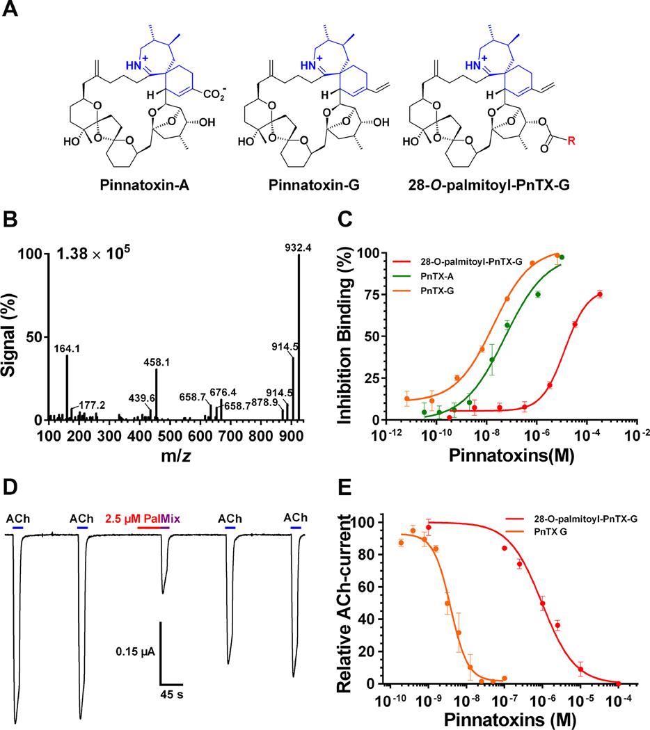
FIGURE 3.
3A. Chemical structure of pinnatoxin-A, pinnatoxin-G and 28-O-palmitoyl pinnatoxin-G; R = ester 16:0. 3B Mass spectrum of 28-O-palmitoyl ester of pinnatoxin-G showing its fragmentation pattern at the collision-induced dissociation condition for MRM. 3C. Inhibition binding potency of 28-O-palmitoyl pinnatoxin-G compared to its precursor molecule pinnatoxin-G and to pinnatoxin-A. Each point represents the mean value ± SEM of three independent experiments using the Microplate-RBAssay. D. Antagonistic effect of 2.5 μM 28-O-palmitoyl ester of pinnatoxin-G on Torpedo-nAChR of muscle embryonic type recorded using two-electrode voltage clamp electrophysiology. The perfusion protocol was as follows: A clamped oocyte at a holding potential of −60 mV was perfused with 25 μM ACh (ACh) for 15 s at 150 s intervals. Thereafter, the oocyte was perfused with 2.5 μM 28-O-palmitoyl ester of pinnatoxin-G (Pal) for 45 s and immediately after it was exposed to a mixture of 25 μM ACh and 2.5 μM 28-O-palmitoyl ester of pinnatoxin-G (Mix). The oocyte was washed with Ringer-Ba and pulses of 25 μM ACh were recorded twice at 150s intervals. E. Concentration-dependent antagonistic effect of 28-O-palmitoyl ester of pinnatoxin-G and pinnatoxin-G on Xenopus laevis oocytes having incorporated Torpedo muscle-type (α1)2βγδ nAChR into their plasma membranes. The peak amplitudes of the ACh-evoked current values (mean ± SEM; 5 oocytes per concentration) were normalized to control ACh-peak amplitudes recorded from the same oocyte to yield fractional response.
Abbreviations: m/z: Mass-to-charge ratio; PnTX-A: Pinnatoxin-A; PnTX-G: Pinnatoxin-G; PnTX-G-palmitate: 28-O-palmitoyl pinnatoxin-G.

