fig 4.

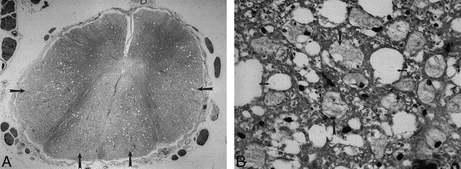
A, Luxol fast blue stain of the spinal cord in a patient with AIDS-associated myelopathy (not from our series) shows pathologic changes predominantly involving the posterior and lateral columns (arrows).
B, A magnified view of the same specimen as in A shows vacuolization (small arrows) and lipid-laden macrophages (large arrows) scattered throughout the involved cord.
(Reprinted with permission from Mandell GL. Atlas of Infectious Diseases, I: AIDS. 2nd ed. Philadelphia: Current Medicine, Inc; 1997.)
