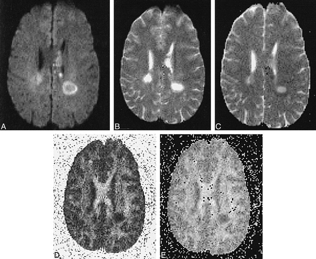
fig 2.

Patient V.Y: 24-year-old man with 7-month history of relapsing-remitting MS presented with quadriparesis, most severe in upper extremities, and left periventricular acute plaque.
A–E, Note delineation of the hyperintense rim on diffusion-weighted image and relative hypointensity of lesion center (A). T2-weighted echo-planar image shows marked central hyperintensity and slightly lower signal in rim (B). Center has increased diffusibility, appearing almost as bright as CSF, and has a low ADC value (dark) in the rim on calculated ADC map (C). FA (D) and eccentricity (E) maps reveal intermediate anisotropic values in rims relative to lesion centers, which approach isotropy. ADC values: NAWM, 0.00071; left periventricular acute plaque center, 0.00136; left periventricular acute plaque rim, 0.00066. FA values: NAWM, 0.430; left periventricular acute plaque center, 0.135; left periventricular acute plaque rim, 0.362.
