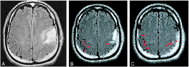
fig 1.

A–C, Assessing the feasibility of surgery in a tumor patient, case 9 (Table 2): 38-year-old man with a biopsy-proved grade 2 oligodendroglioma in the left perirolandic region (A). Functional MR imaging was performed to assess the anatomic relationship between the tumor and the functional hand area. The thresholded activation map (red pixels) was superimposed on a FLAIR image. B represents the most inferior section on which functional activation was identified and C represents a more cephalic section. The apex of the tumor was in contact with the most inferior portion of the functional MR imaging activation (B). After considering the risk versus benefit of a proposed gross total resection, the patient opted for treatment with external beam radiation and no further surgery
