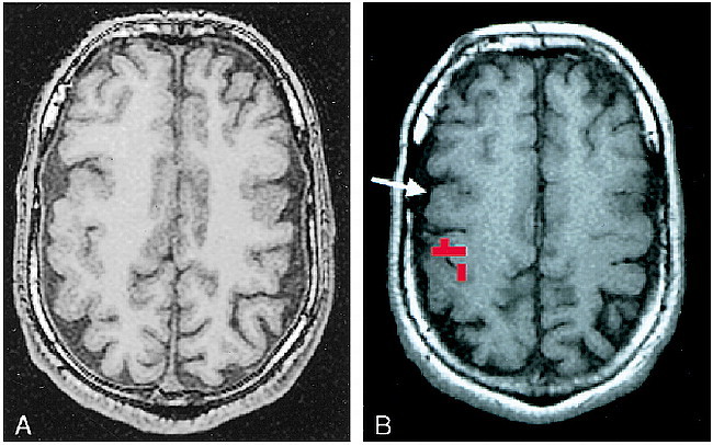
fig 2.

A and B, Assessing the feasibility of surgery in an epilepsy patient, case 12 (Table 1): 31-year-old man with life-long medically intractable seizures. An anatomic MR study (A) revealed bifrontal cortical developmental anomalies. Prolonged video-EEG recording showed unequivocally that the patient's typical seizures arose from the right frontal area. The characteristic landmarks used to define the anatomic central sulcus were absent. Functional MR imaging was performed (B) to ascertain the topographic relationship between the right frontal developmental anomaly and the hand sensorimotor functional area. This study showed that the functional hand area was posterior and superior to the anatomic developmental anomaly (arrow). The anatomic abnormality was resected, and the patient has been seizure-free for over 2 years
