fig 1.

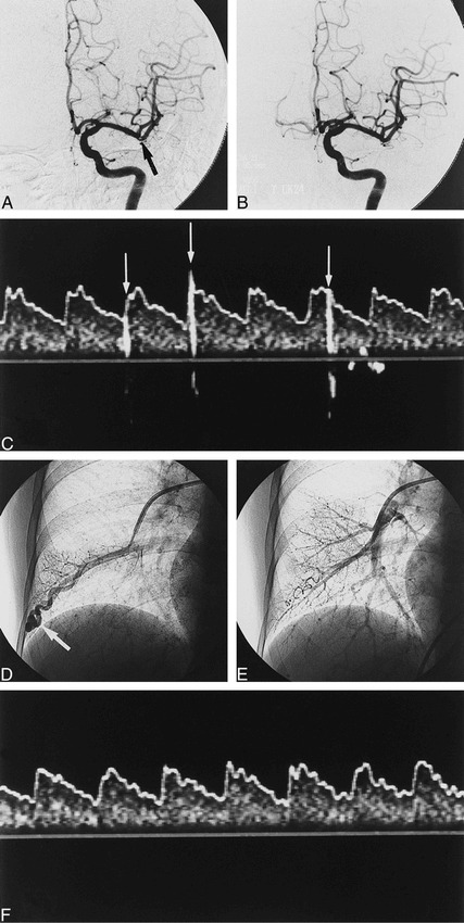
Images from the case of a 62-year-old right-handed woman who experienced the sudden onset of aphasia and right-sided hemiparesis 1 hour after the onset of stroke.
A, Cerebral angiogram shows the intraluminal filling defect (arrow) at an ascending branch of the left MCA 2 hours after the onset of symptoms.
B, After 240,000 IU of urokinase was administered locally through a microcatheter, the filling defect disappeared.
C, Administered before embolization therapy of the PAVF, TCD with saline contrast medium during normal breathing shows many high-intensity transient signals (arrows) from the left MCA 5 days after the onset of stroke.
D, Selective pulmonary angiography shows a PAVF (arrow) in the right lower lobe.
E, After embolization therapy with metallic coil, the feeding vessel to the PAVF is completely occluded.
F, TCD with saline contrast medium failed to reveal any HITS immediately after embolization therapy of the PAVF.
