fig 1.

Case 1.
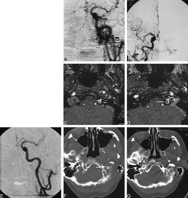
A, Anteroposterior view of left common carotid artery demonstrates a DAVF at skull base. Fistula was located in superior aspect of dilated anterior condylar vein (arrow), medial to jugular bulb (curved arrow).
B, Anteroposterior view of right common carotid artery demonstrates fistula (arrow) medial to jugular bulb. This view was used to direct transvenous embolization to exact site of the fistula.
C, MR angiography source image from three-dimensional time-of-flight sequence (54/9/1 [TR/TE/excitations]) at level of hypoglossal canal shows abnormal flow signal in left hypoglossal canal (arrow).
D, MR angiography source image from three-dimensional time-of-flight sequence (54/9/1) at level of junction of sigmoid sinus and jugular bulb demonstrates abnormal intraosseous flow (arrow) medial to jugular bulb.
E, Anteroposterior view of right common carotid artery following transvenous coil embolization confirms occlusion of fistula.
F, CT scan following transvenous embolization demonstrates portion of coil mass in hypoglossal canal (arrow).
G, CT scan after embolization shows coil mass to be partially intraosseous and located medial to jugular bulb.
