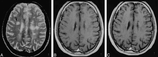
fig 2.

A–C, T1 isointense nonenhancing lesion in the right frontal white matter. Axial T2-weighted image (2200/80/1) (A) and contrast-enhanced T1-weighted image (550/14/2) (B) at a comparable anatomic level are shown at day 0 and at 12 months (C). The lesion (arrow, A) shows no significant change on the 12-month image, and the MTR values remained between 35% on the baseline image and 33% on the 12-month image.
