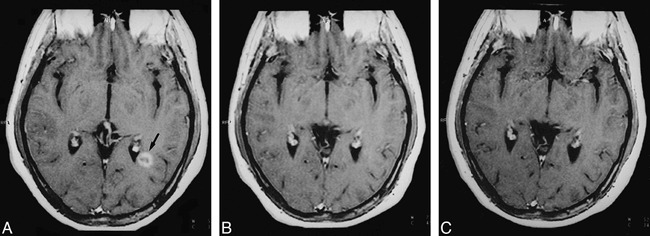
fig 3.

A–C, Serial MR follow-up of a ring-enhancing lesion in the left periventricular temporal white matter. The lesion (arrow, A) is shown at three time points: baseline (A), 1 month (B), and 12 months (C) on contrast-enhanced T1-weighted images (550/14/2). The ring-enhancing pattern at baseline disappears on the 1-month image. At 12 months, the lesion is completely isointense with respect to NAWM on contrast-enhanced T1-weighted images. In this lesion, the MTR increased from 22% on the baseline image to 36% after 12 months.
