
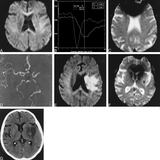
fig. 2. 81-year-old woman scanned 2 and 19 hours post ictus.
A, Diffusion-weighted imaging at 2 hours shows subtle restricted diffusion in the left deep cerebral structures, not appreciated prospectively.
B and C, Perfusion-weighted imaging bolus-tracking curves (B) for selected regions (C) show approximately 6-second delayed transit to the distal left middle cerebral artery vessels (solid line).
D, MR angiography demonstrates decreased flow-related enhancement in the left middle cerebral artery territory.
E, Diffusion-weighted imaging at 19 hours reveals acute infarct in left basal ganglia and insular cortex.
F, T2*-weighted imaging demonstrates a new 1-cm focus of profound hypointensity in the left putamen, consistent with focal hemorrhage or localized desaturation of hemoglobin.
G, CT scan at 2 days reveals a 1-cm focus of petechial hemorrhage in the left putamen surrounded by ischemic infarct.