
fig 4.
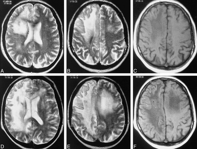
Marked progression of PML documented by serial MR studies.
A and B, Axial T2-weighted images (3500/95/1) show the right frontal lobe confluent hyperintense signal abnormalities extending from the periventricular white matter to the subcortical white matter, with much milder white matter involvement in the right parietal lobe and minimal involvement of the left cerebral hemisphere.
C, Axial T1-weighted image (600/15/1) shows corresponding low signal abnormalities in the affected white matter on the right as well as minimal mass effect on cortical sulci.
D–F, Eight weeks later, marked progression of disease is evident with extension and increasing confluence of the right frontal and parietal lobe lesions, corpus callosum involvement, and greater involvement of the left cerebral hemisphere. Also seen is an increase in white matter low signal abnormality on axial T1-weighted image (600/15/1). Patient died 7 days after this study. (Biopsy tract is also evident in the right cerebral hemisphere.)