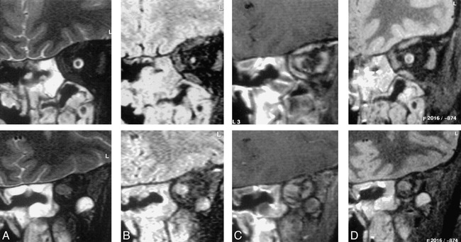
fig 2.

Coronal images through the orbits of a patient with an orbital apex meningioma. The sequences are T2-weighted SPIR (A), SPIR/FLAIR (B ), T1-weighted SPIR with contrast (C ), and STIR (D).
Images in the mid orbit (top row) show normal appearances on the STIR (top of D) and T2-weighted SPIR (top of A) images. The SPIR/FLAIR image (top of B) clearly shows increased signal in the optic nerve itself. Postcontrast T1-weighted SPIR (top of C) shows thickening of the optic nerve sheath because of meningioma en plaque. Images at the orbital apex (bottom row) demonstrate a mass lesion in the position of the optic nerve–sheath complex. The contrast between the lesion and surrounding tissues is greater on the SPIR/FLAIR image (bottom of B) than on the T2-weighted SPIR (bottom of A) or STIR (bottom of D) images. On the postcontrast T1-weighted SPIR image (bottom of C) the lesion shows inhomogeneous enhancement and is difficult to distinguish from adjacent enhancing extraocular muscles.
