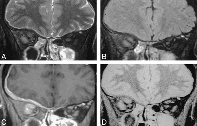
fig 4.

Coronal images through the orbit in a patient with an orbital apex meningioma. The extensive intracranial en plaque spread is shown well on the T1-weighted SPIR sequence with contrast (C ) and also can be appreciated on the T2-weighted SPIR (A) and STIR (D) images it is outlined by high-signal CSF. Although the meningeal thickening can be seen on the SPIR/FLAIR sequences (B ), its presence and extent were not appreciated by either of the radiologists who were reporting on this scan in isolation.
