fig 1.

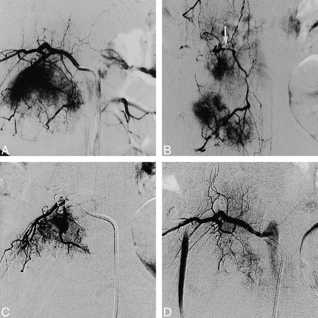
Case 7: 27-year-old woman with a giant cell tumor in the L3 vertebral body who underwent embolization 2 days before surgery.
A, Preembolization angiogram of the right second lumbar artery shows hypervascular supply to the tumor.
B, Selective injection of the dorsispinal artery at L2 (arrow) shows the extraspinal longitudinal anastomoses from L1 to L4 with tumor supply by L2 and L3.
C, Superselective injection of the feeding artery near the dorsispinal branch was followed by infusion of PVA particles.
D, Postembolization angiogram shows nearly total occlusion of the feeding artery and patency of the normal branches.
