fig 2.

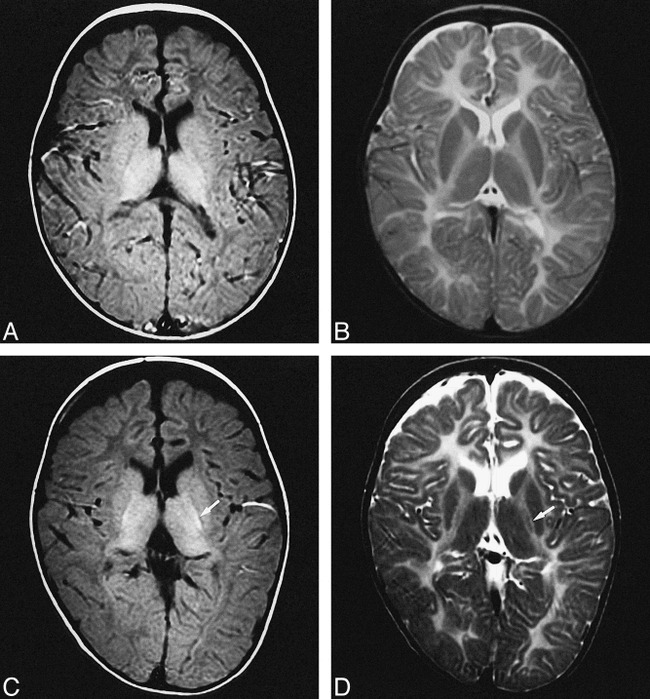
MR images of patient 4 with PMD attributed to PLP duplication (connatal form).
A and B, T1- and T2-weighted images obtained when the patient was 1 year 9 months old.
C and D, T1- and T2-weighted images obtained when the patient was 5 years old. Myelination extends into the internal capsule over a 3-year period (white arrow).
