fig 6.

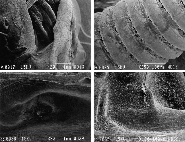
Scanning electron microscopic appearance of orifice of embolized aneurysms.
A and B, Lower (×20) and higher (×250) magnification of orifice treated with standard GDCs. Surface of coils were exposed to the arterial lumen and only fibrin/leukocyte complex was seen on surface.
C and D, Lower (×23) and higher (×100) magnification of orifice treated with laminin GDC-Is. Orifice was covered completely with neoendothelial cell layer.
