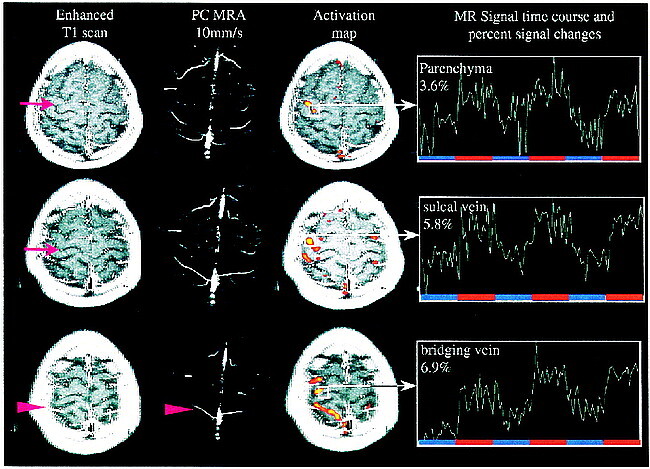
fig 2.

MR signal time course correlation with contrast-enhanced T1-weighted scans and phase-contrast MR angiography (S5). Contrast-enhanced studies reliably show sulcal and superficial veins, whereas the phase-contrast MR angiography with given flow sensitivity demonstrates vessels the size of large bridging veins. Significant change in MR signal (ΔS) is seen in bridging veins even close to superior sagittal sinus. From this data, one can presume that dilution of increased oxyhemoglobin content and therefore decay of ΔS takes place when bridging veins enter large draining sinuses. Arrows point at sulcal veins, arrowheads at large bridging veins.
