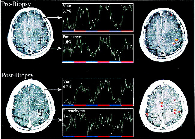
fig 4.

Functional MR and MR signal time course. This patient was evaluated for presurgical planning of a space-occupying lesion near central sulcus (Astrocytoma III) (S7). The first fMR imaging study shows dual activation in slice of interest with parenchymal activation located in close proximity to tumor. A stereotactic biopsy was performed after which the patient suffered from transient mild paresis of the small right hand muscles. A postbiopsy MR image demonstrates that the biopsy involved a previously activated region posterior to the lesion. Functional study again shows dual activation with parenchymal activation close to biopsy tract and lateral venous activity unaffected by biopsy. This case illustrates the importance of differentiating task-related hemodynamic changes of small parenchymal venules and large draining veins.
