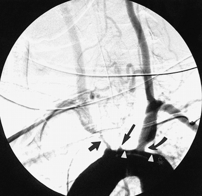
fig 5.

Oblique view of the aortic arch during aortography shows thickening and flattening of the superior wall of the aortic arch (arrowheads). There is no dissection, but study confirms total occlusion at the origin of the left common carotid artery (long arrow), moderate stenosis of the left subclavian artery (curved arrow), and a tight stenosis at the origin of the right brachiocephalic artery (short arrow).
