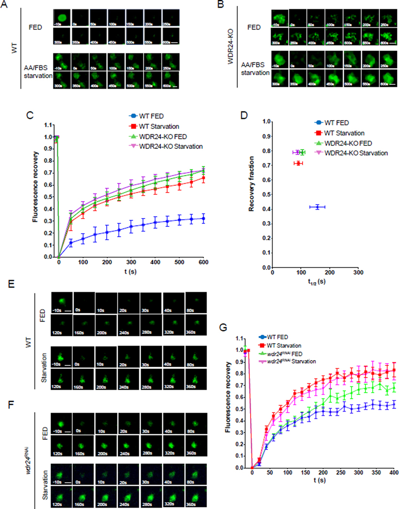
Figure 2. The GATOR2 complex inhibits the recruitment of TSC to lysosomes.
(A) Time point images from the Halo-TSC2 FRAP experiment in WT HeLa cells. The 0 s image represents photobleaching. Scale bar: 0.5 μm.
(B) Time point images from the Halo-TSC2 FRAP experiment in WDR24-KO cells.
(C) Fluorescence recovery versus time curves in A and B. A total of 30 lysosomes from different cells were used to plot the curves for each treatment. Error bars represent standard error.
(D) Plot showing the relation between the recovery fraction versus half time (t1/2) from curves in C. Error bars represents standard error.
(E) Time point pictures from the GFP-TSC1 FRAP experiment in WT Drosophila ovary.
(F) Time point pictures from the GFP-TSC1 FRAP experiment in wdr24RNAi Drosophila ovary. Scale bar: 2 μm.
(G) Fluorescence recovery versus time curves in E and F. A total of 10 lysosomes in different ovaries from each treatment were used in plotting the curve. Error bars represent standard error. See also Figure S2, S3 and S4.

