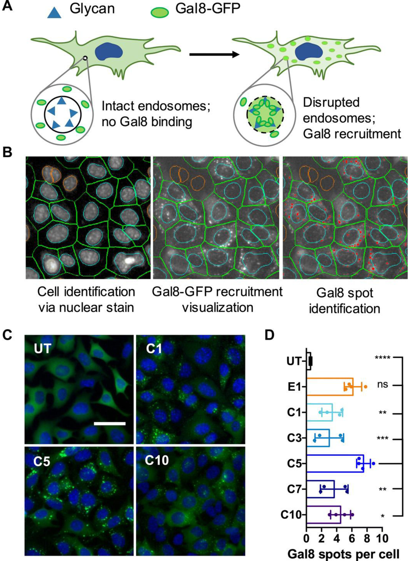
Figure 4.

A Gal8 recruitment assay shows PBAE nanoparticle-mediated endosomal disruption. (A) Gal8-GFP is dispersed throughout cells with intact endosomes. In disrupted endosomes, Gal8-GFP binds to endosomal glycans, resulting in punctate fluorescent dots. (B) Image-based analysis of acquired microscope images as shown in (C) can be used to quantify endosomal disruption. Individual cells are identified through nuclear staining (left); Gal8-GFP recruitment is visualized in the green fluorescence channel (middle); and punctate GFP+ spots are identified and counted (red dots). (C) Representative images of Gal8-GFP+ B16 cells left untreated (UT) or treated with carboxylated PBAE/BSA nanoparticles, where C1, C5, and C10 represent particles made using different polymers (scale bar: 50 μm). (D) Endosomal disruption is quantified by the number of Gal8-GFP spots per cell following carboxylated nanoparticle treatment. Particles made from different polymers are listed on the y-axis, where UT is an untreated group. From [54]. Reproduced with permission from American Association for the Advancement of Science (AAAS).
