Figure 7.

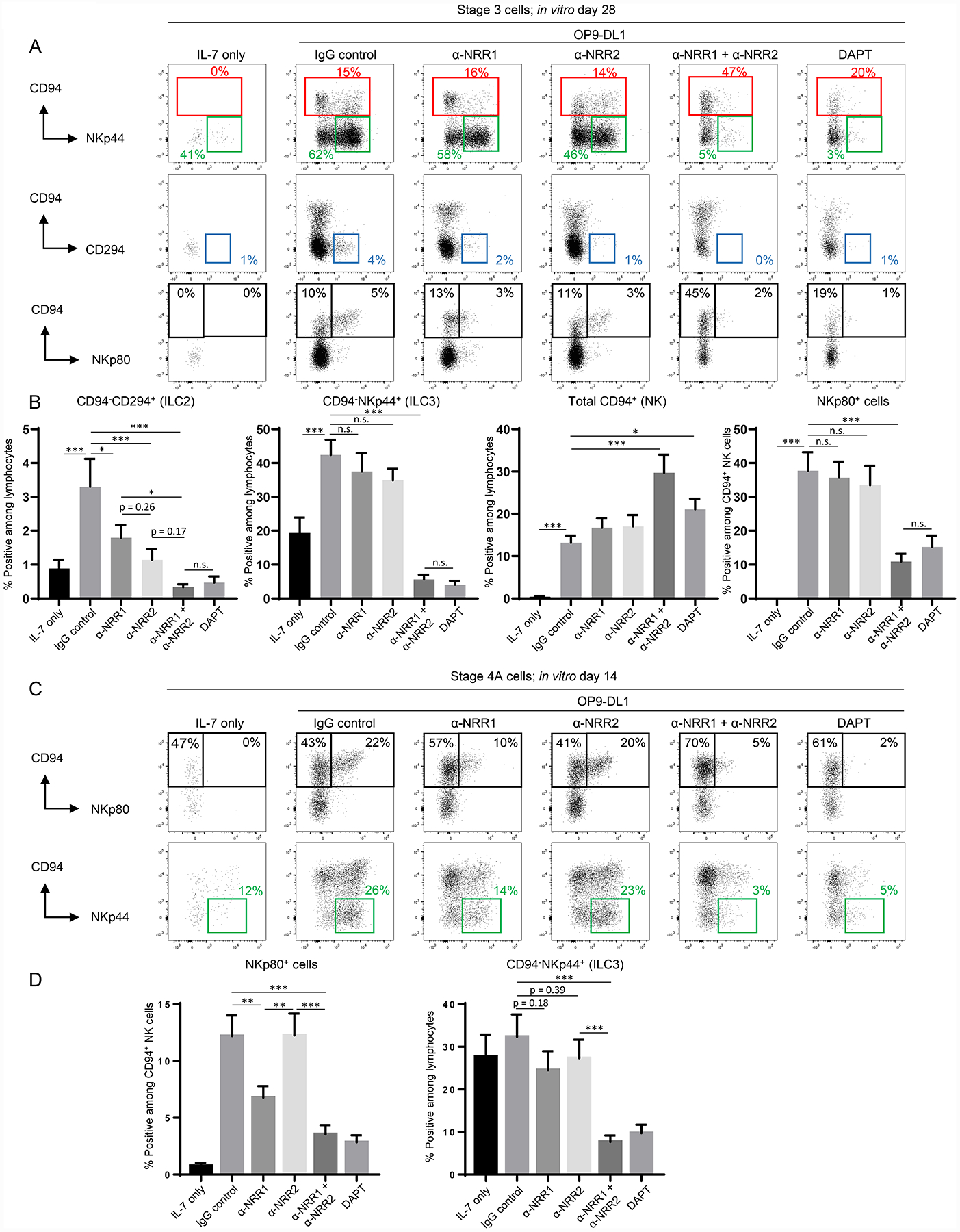
Stage-specific effects of NOTCH1 and NOTCH2 on ILC development. (A) Representative (n = 13; 4 independent experiments) surface flow cytometry analyses of ILCs generated in vitro following 28 day culture of freshly purified tonsil-derived stage 3 cells with IL-7 alone or IL-7 + OP9-DL1 cells with the addition of IgG control antibody, anti-NRR1 and/or anti-NRR2 (5 μg/ml each), or DAPT. (B) Quantification of CD94−CD294+ (ILC2s), CD94-NKp44+ (ILC3s), total CD94+ (NK), and NKp80+ cells generated in vitro following 28 day culture of freshly purified tonsil-derived stage 3 cells in the conditions described in (A). Data are represented as mean ± SEM. n.s. = not significant (p > 0.05); * p < 0.05; ** p < 0.01; *** p < 0.001. (C) Representative (n = 18; 6 independent experiments) surface flow cytometry of ILCs generated in vitro following 14 day culture of freshly purified tonsil-derived stage 4A cells with IL-7 alone or IL-7 + OP9-DL1 in the presence of IgG control, anti-NRR1 and/or anti-NRR2, or DAPT. (D) Quantification of NKp80+ and CD94−NKp44+ cells generated in vitro following 28 day culture of freshly purified tonsil-derived stage 4A cells in the same conditions described in (C). Data are represented as mean ± SEM. ** p < 0.01; *** p < 0.001.
