
Figure 1:
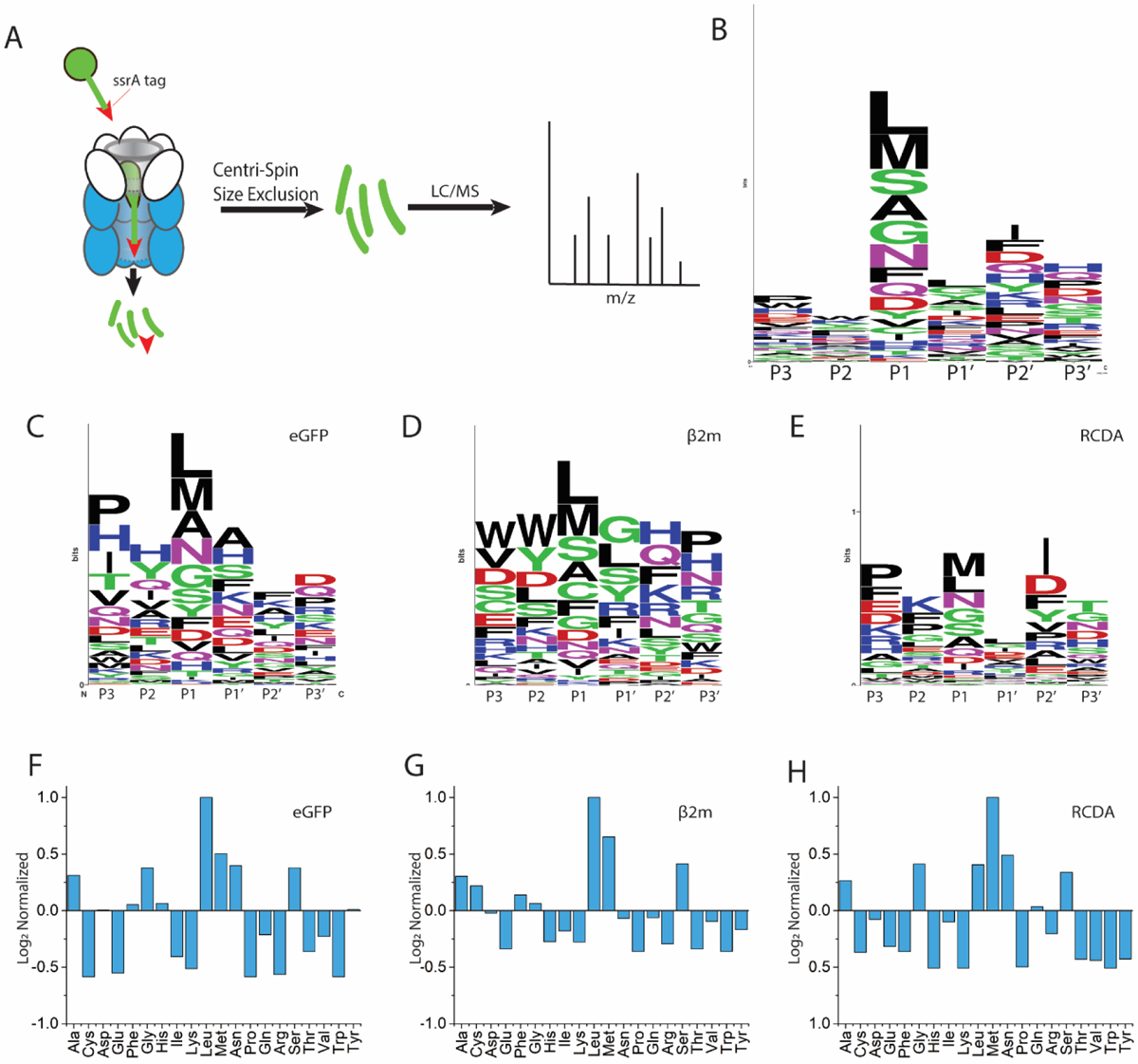
Primary structure cleavage specificity of ClpXP. (A) Schematic depiction of tagged protein digestion, separation, and analysis. Residues directly adjacent to the cleavage site in the N-terminal direction are indicated as P1, P2 and P3, and residues directly adjacent to the cleavage site in the c-terminal direction are indicated as P1’, P2’ and P3’, numerically increasing as they are further from the cleavage site. Weblogo representation of cleavage preferences for all substrates (B), GFP (C), reduced β2m (D), and RcdA (E). Amino acid preferences at the P1 position normalized to presence in the given protein for GFP (F), reduced β2m (G), and RcdA (H).