Fig. 2.
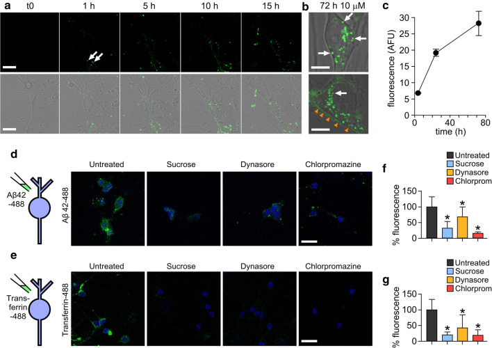
Alexa fluor-488 labelled Aβ42 oligomers are internalised into primary neurons by a dynamin-dependent mechanism. a Confocal images showing one 0.5 μm z-slice from the middle of the cell body of neurons treated with 10 μM Aβ42 oligomers over 15 h. Representative images from three experiments. Oligomers can be seen associating with the plasma membrane (white arrows) and internalising into the cell. Scale bar is 25 μm. b Magnified example images of cell bodies after 72 h containing Aβ42-488 oligomers in vesicles within the cytoplasm (white arrows) and along the plasma membrane (orange arrowheads). Representative images from four experiments. One 0.5-μm z-slice is shown from the middle of the cell body. Scale bar is 10 μm. c Increase in Alexa Fluor 488 fluorescence intensity within the cell body over time. n (cells) 4 h = 14, 24 h = 31, 72 h = 11. Plot shows mean ± SEM. Inhibition of uptake of Aβ42-488 oligomers (d) or transferrin-488 (e) after 30 min incubation with 0.5 M sucrose, 10 μM dynasore or 25 μg/mL chlorpromazine. Neuronal nuclei stained with DAPI (4′,6-diamidino-2-phenylindole) are shown in blue and Aβ42 or transferrin are green. One 0.5-μm z-slice is shown from the middle of the cell body. Scale bar is 25 μm. f, g Quantification of 488 fluorescence intensity from the cell body from three experiments normalised to untreated. A Kruskal–Wallis ANOVA was carried out followed by pairwise comparisons using Dunn’s test and all were significant compared to untreated (*p < 0.05)

