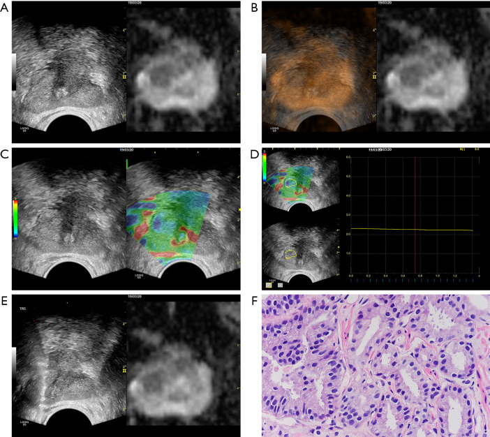
Figure 1.
A 60-year-old patient (PSA =6.09 ng/mL, fPSA =0.48 ng/mL, fPSA/PSA =0.079, TRUS-derived prostate volume =20.8 cm3, PSAD =0.29 ng/mL/cm3) had a pre-biopsy MRI that detected an abnormal signal area of about 14 mm × 10 mm at the right transition zone of the prostate; the PI-RADS score was 4. (A) Image registration of ADC-US fusion; (B) the overlay image of the ADC-US fusion; (C) elastography detected the suspicious zone with increased stiffness, which was previously detected by MRI; (D) the EQS of the suspicious zone was 2.3; (E) targeted biopsy of the lesion detected by MRI and elastography; (F) targeted biopsy result was positive for PCa (HE staining, ×40), with a GS of 7 (3+4) and classified as group 2 according to the WHO/ISUP classification system.

