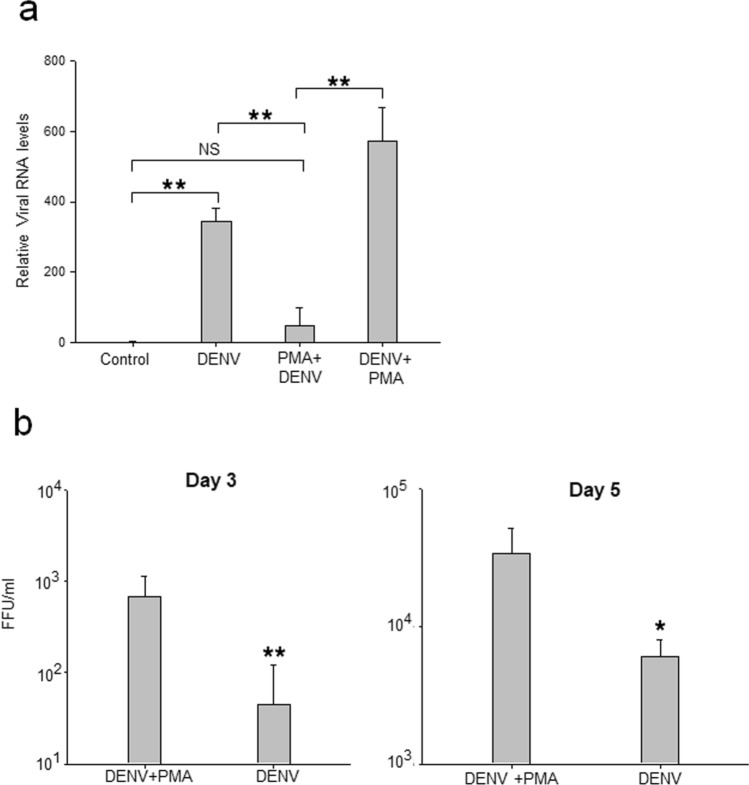
Figure 2.
DENV infection of MEG-01 cells. MEG-01 cells were mock-infected, or infected with DENV at a multiplicity of infection (MOI) 1, and the cells and culture supernatant were harvested on different days post-infection (pi). In parallel, cells were pre-treated with PMA 2 h before infection (PMA + DENV) or infected with DENV followed by PMA treatment (DENV + PMA). (a) The total RNA from cells was isolated on day 2 pi and relative levels of viral RNA determined by qRT-PCR using Gapdh transcripts for normalization. (b) The culture supernatants from virus-infected cells were collected on days 3 and 5 pi and DENV titers determined by FFU assays. All the experiments were done more than 3 times and data represent the mean ± standard deviation. Statistical analyses were done using the one-way analysis of variance (ANOVA). *p < 0.05 and **p < 0.005.

