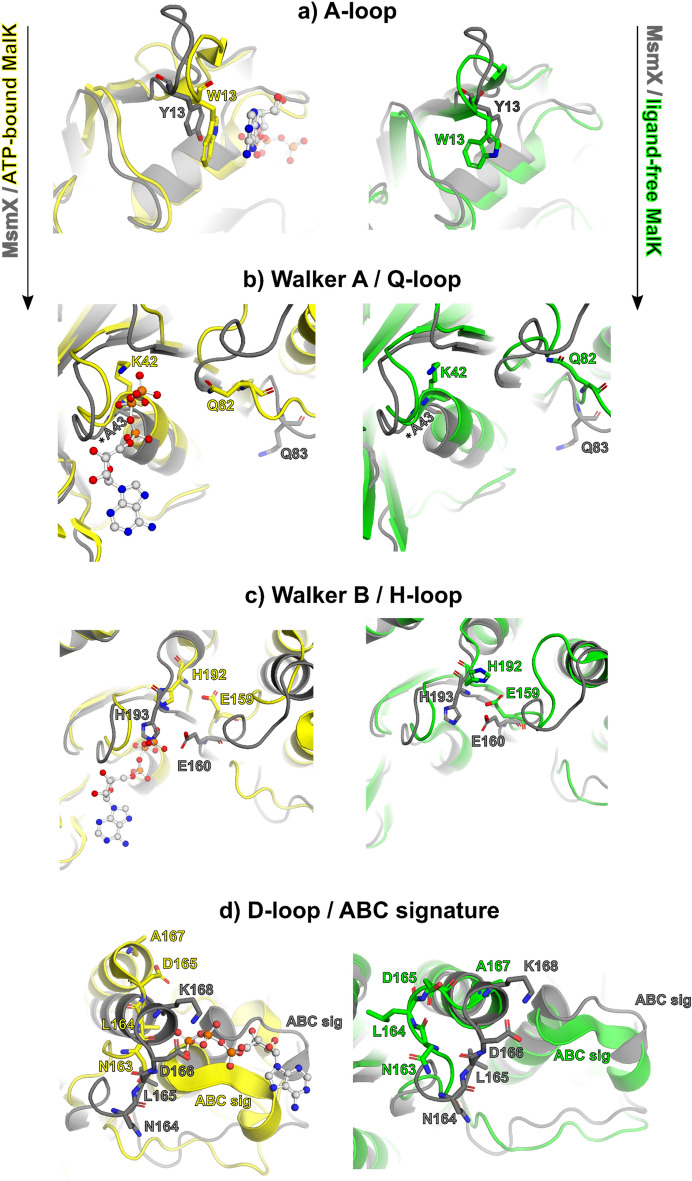
Figure 8.
Structural characterization of MsmX functional motifs. Representation of NBDs functional motifs of the structural superposition of MsmX K43A (grey), ligand-free MalK (green) (PDB ID 1Q1E:A) and ATP-bound MalK (yellow) (PDB ID 1Q12:A): A-loop (a), Walker A and Q-loop (b), Walker B and H-loop (c), D-loop and ABC signature (d). The most important molecular determinants in each functional motif are represented as sticks and labelled with the color correspondent to the structure. The ATP ligand from PDB ID 1Q1E is represented as ball and stick. Figures obtained with The PyMOL Molecular Graphics System, Version 2.0.5 Schrodinger, LLC (https://pymol.org/2/).

