Figure 1.
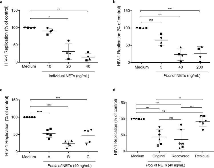
NETs inhibit HIV-1 production in macrophages. (a) Macrophages from healthy donors were infected in vitro and treated with NETs obtained from different individuals for 3 h, washed and maintained in culture (n = 4). (b) HIV-1-infected macrophages were treated with different concentrations of one pool of NETs during 3 h, washed and maintained in culture (n = 4). (c) HIV-1-infected macrophages were treated with three different pools of NETs during 3 h, washed and maintained in culture (n = 5). (d) One pool of five NETs was centrifuged and recovered using Centricon tubes of 100 kDa; thus, HIV-1-infected macrophages were treated with the original or with the recovered pool, or with the residual supernatant during 3 h; cells were washed and maintained in culture. In all these assays, HIV-1 replication was measured 12–14 days post-infection by quantifying the levels of HIV-1 p24 antigen in culture supernatants by ELISA. Data were analyzed with the Kruskal–Wallis test, with Dunn's post-test (a,b) and One Way ANOVA, with Turkey post-test (c,d). Each point represents a donor. Data represent means ± SD. Viral production in controls (cells incubated only with medium) in ng/mL: (a) = 13.8 ± 5.9; (b) = 17.5 ± 10.1; (c) = 14.7 ± 8.5; (d) = 13.06 ± 9.6. *p < 0.05; **p < 0.01; ***p < 0.001; ****p < 0.0001. ns non-significant.

