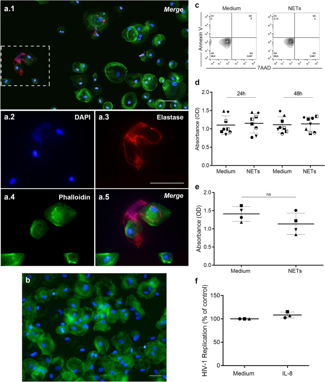
Figure 2.
NETs interact with macrophages without affecting cell viability. (a) Interaction of NETs with macrophages from HIV-1 infected cultures. HIV-1-infected macrophages were exposed to NETs for 30 min and, after fixation, NETs were labeled with DAPI and anti-NE antibodies, while cells were visualized by phalloidin and DAPI labeling. Images (a.2) to (a.5) depict digital amplification of the area delimited by the dotted lines in (a.1), highlighting NET structures around the cells. Representative images of four different assays. Scale bar = 50 μm. (b) Removal of NETs by washing macrophages culture. Macrophages were incubated with NETs during 3 h, washed, fixed and labeled as described above. Observe that NET components were not detected. Representative image of three different assays, obtained after detailed and careful analysis of several microscopic fields by two independent investigators. Scale bar = 100 μm. (c) Viability of macrophages exposed to NETs. Macrophages were treated for 3 h with NETs, washed, and cell viability was evaluated 24 h later by flow cytometry (representative image of 2 independent experiments). Additionally (d), cell viability was assessed by XTT method after 24 h and 48 h or (e) in macrophages that had not been washed 14 days after NET treatment. ns non-significant. (f) HIV-1-infected macrophages were treated with IL-8 at a concentration equivalent to that carried over by the induced NETs, and no changes in HIV-1 production were observed in this condition.

