Figure 7.
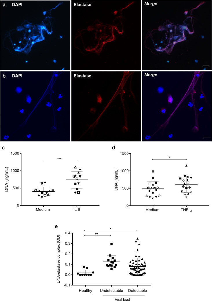
Neutrophils from HIV-1 patients release NETs, which are present in patients’ plasma. (a) Neutrophils from HIV-1 patients or (b) from healthy donors were activated with IL-8 (50 ng/mL) for 3 h at 37 °C, fixed and labeled with DAPI and anti-NE antibodies. Merged images show the colocalization of the two markers and the network morphology of NETs. Images are representative of at least 6 (HIV-1 patients) or 4 (healthy donors) assays. Scale bar, 10 µm. (c,d) Neutrophil from HIV-1 patients were stimulated with IL-8 (50 ng/mL; n = 12) or TNF-α (20 ng/mL; n = 17) and NET release was quantified with the Picogreen dsDNA kit. Each point represents a donor. Data were analyzed using paired Student’s t test. (e) Quantification of DNA-elastase complexes in the plasma of HIV-1-infected patients. Data were analyzed using ANOVA. Viral loads in HIV-1 RNA copies/mL: Undetectable: < 40; Detectable: > 40. *p < 0.05; **p < 0.01; ***p < 0.001.

