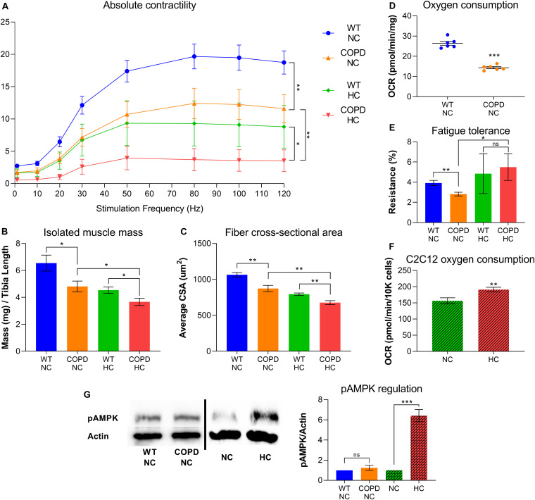
FIGURE 1.
COPD and hypercapnia synergistically contribute to muscle atrophy, but they are antagonistic processes regarding fibers respiratory capacity. (A) Isolated EDL muscle contractility indicates that murine COPD (orange line) causes reduced absolute force-generation compared with wild type (WT) animal (blue line), which is incremented by hypercapnia (green line) and even further by the combination of hypercapnia and murine COPD (red line). Absolute force-generation capacity reflects muscle work dependent on muscle mass (n = 4). (B) Isolated EDL muscle mass and (C) fibers average cross-sectional area (CSA) follow the same incremental pattern of COPD and hypercapnia described in (A) (n = 4). (D) Oxygen consumption rate (OCR) by plate respirometry obtained with Seahorse§ technology demonstrates that EDL muscle reduced respiratory capacity induced by normocapnic murine COPD (n = 6). (E) Fatigue-tolerance, which partially depends on the fibers oxidative capacity, is reduced in normocapnic (NC) murine COPD versus wild type (NC-WT) animal (n = 4). However, the exposure of these animals to hypercapnic (HC) conditions leads to an improvement of their fatigue-tolerance (n = 4). (F) OCR by plate respirometry obtained with Seahorse§ technology indicates that C2C12 cells grown for 48 h in hypercapnic conditions demonstrate elevated respiratory capacity (n = 7). (G) Activation of AMPK, as reflected by its phosphorylation at threonine 172 (pAMPK) is not evident in the COPD animal but robustly demonstrated by muscles from animals exposed to hypercapnia (n = 4), *p < 0.05, **p < 0.01, ***p < 0.001. For technical details about the methods, see our recent publications (Jaitovich et al., 2015; Balnis et al., 2020a,b,c; Korponay et al., 2020). Presented data is original and thus has not been previously reported.

