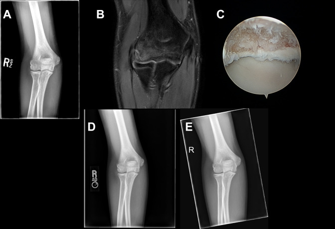
Figure 1.
(A) Preoperative anteroposterior radiograph and (B) selected T2 coronal magnetic resonance image of a 14-year-old male (patient H) with a right capitellar osteochondral lesion who underwent arthroscopic loose body removal and microfracture. (C) Arthroscopic image demonstrates the osteochondral defect prior to microfracture. (D) Three-month and (E) 6-month postoperative anteroposterior radiographs show ossification of bone within the region of microfracture with healing, including resolution of lucency, sclerosis, and restoration of articular contour. Evidence of healing is present 3 months postoperatively.

