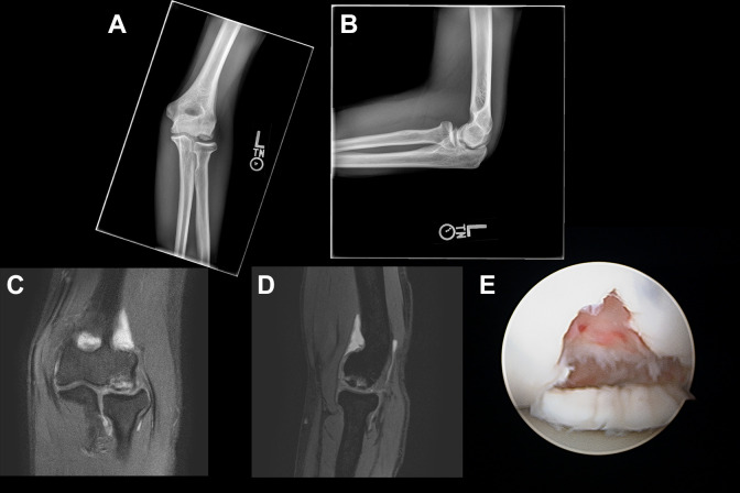
Figure 3.
(A) Anteroposterior and (B) lateral radiographs of the left elbow of a 16-year-old female gymnast (patient A) who sustained a repeat injury from continued gymnastics 2 years after arthroscopy, chondroplasty, microfracture, and loose body removal and required an OATS procedure. After the index procedure, there was restoration of the articular contour and sclerosis indicative of radiographic healing 3 months postoperatively. These radiographs are from just before the OATS procedure and illustrate the new large capitellar osteochondral defect that did not resolve with nonoperative management. Selected T2 (C) coronal and (D) sagittal magnetic resonance images of the left elbow. Imaging was obtained before the OATS procedure and demonstrates an unstable osteochondral fragment. (E) Arthroscopic image shows the large capitellar osteochondral defect after repeat injury. After the OATS procedure, the patient had clinical and radiographic healing and was able to return to full activities, including gymnastics. OATS, osteochondral transfer system with allograft.

