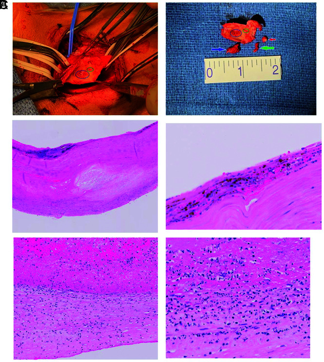
FIG 2.
Gross and microscopic pathology. A, Surgical view in patient 2. The blue circle indicates the area to which the thrombus was adherent, and the green circle indicates the area to which the contiguous portion of the thrombus was adherent. Arrows represent the larger portion of thrombus (blue), smaller portion of thrombus (green), and calcified plaque at the external carotid origin (red). B, Microscopic examination of a specimen from patient 2. Hematoxylin-eosin stain shows intimal thickening, plaque formation, and calcification. Higher magnification shows an area of intima with mononuclear inflammatory cells and evolving plaque formation. C, Microscopic examination of a specimen from patient 1. Low-power hematoxylin-eosin stain shows the thrombus adherent to the wall (upper portion is thrombus; lower portion is the wall). Higher-power view of an area of adherent thrombus shows the intima with inflammatory infiltrate as well as some degenerating cellular debris that may indicate apoptosis.

