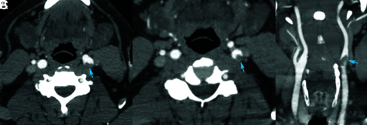
Fig 2.
Axial (A and B) and coronal (C) images from a CT angiogram in a patient presenting with left MCA syndrome and a left middle cerebral artery M1 occlusion. There are eccentric filling defects within the left common carotid artery bifurcation extending into the left internal carotid artery. There are eccentric filling defects within the left common carotid artery bifurcation extending into the left internal carotid artery (blue arrows). Findings are suggestive of acute thrombus.

