Figure 1.
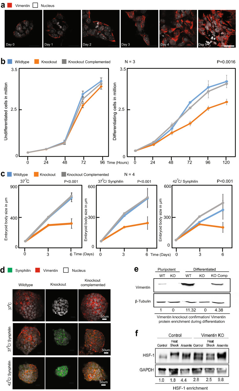
Vimentin Knockout cells are defective in growth and differentiation. (a) mESCs differentiated by the addition of retinoic acid (1 µg/ml) and fixed every 24 h. Immuno-fluorescence for vimentin (red) and nucleus (white) is shown at each time point. Increase in the expression of vimentin was observed at each time point. Images were acquired and processed using NIS elements software (version 3.2) (b) Growth curve comparison between (1) Wildtype, (2) Knockout and (3) Knockout complemented with full-length vimentin in cells maintained in stem cell (2i) media and retinoic acid (differentiation) media. When the cells are maintained in 2i media, no significant change in the proliferation was observed. During differentiation, the wildtype and knockout complemented cells have higher proliferating potential than the knockout cells. The error bars represent standard deviation, statistics were performed by two tailed t-test (P = 0.001).(c) Pluripotent stem cells were seeded on to non-adherent plates without the pluripotency factors leading to the formation of embryoid bodies (EBs). Sizes of the EBs were compared between (1) Wildtype, (2) Knockout and (3) Knockout complemented with full-length vimentin. The sizes were measured every 3 days. The wildtype and knockout complemented cells form larger EBs then the vimentin knockout cells. The experiment was repeated 4 times with measuring 25 embryoid bodies. The same experiment was repeated for cells—(1) with the overexpression of the misfolded protein synphilin and (2) overexpression of the misfolded protein synphilin coupled with heat stress. The error bars represent standard deviation, (P < 0.001). Statistics were performed using two tailed student t-test. (d) Representative images of EBs formed by (1) Wildtype, (2) Knockout and (3) Knockout complemented with full-length vimentin cells. The Vimentin KO was complemented with full length Vimentin-RFP. (e) Western blot for Vimentin for both pluripotent and differentiated (with retinoic acid 1 μg/ml), cells were performed. (f) Western blot for HSF-1 protein after 4 days of differentiation with retinoic acid (1 μg/ml) in wildtype and vimentin knockout cells in two conditions—(1) No stress (2) Heat stress (42 °C).

