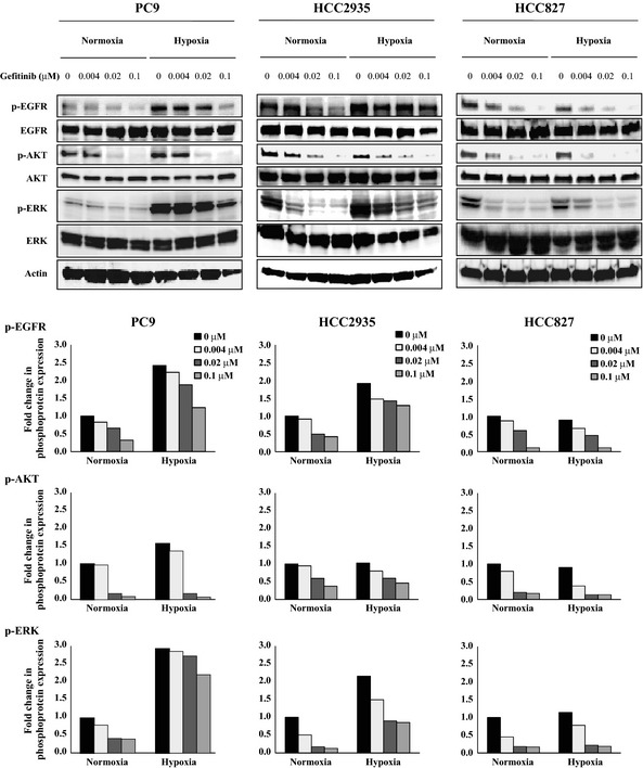
Figure 3.

Effects of gefitinib treatment on the levels of phosphorylated epidermal growth factor receptor (EGFR), AKT, and ERK (p‐EGFR, p‐AKT, and p‐ERK, respectively) under normoxia and hypoxia. The PC9, HCC2935, and HCC827 non‐small‐cell lung carcinoma cells were incubated under hypoxia or normoxia for 48 h, then either left untreated or treated with the indicated concentrations of gefitinib for 3 h. After treatment, the cells were lysed and equal amounts of cell lysates were subjected to Western blot analysis using antibodies against total and phosphorylated EGFR (Y‐1068), AKT, and ERK. The levels of actin served as internal controls for equal protein loading in each lane. The bar graph below each blot shows the fold change in phospho‐protein expression due to treatment with the indicated concentrations of gefitinib. The fold changes were calculated by setting the ratios of the phospho‐protein/total protein band intensities for the untreated normoxia cells to unity.
