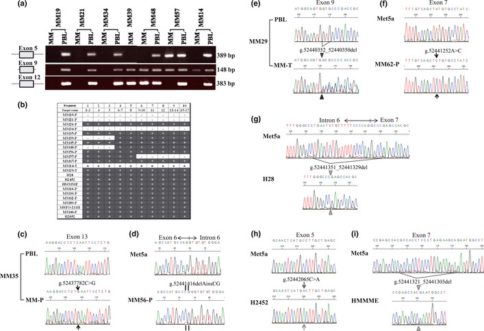
Figure 1.

Genomic alterations of the BAP1 gene. (a) Agarose gel electrophoresis of PCR fragments amplified with the primers detecting exon 5 (fragment 3 in Table 1), exon 9, and exon 12 of BAP1 (fragment 8) from patients with malignant mesothelioma (MM19, MM21, MM34, MM39, MM48, MM57, and MM14). MM, tumor sample of malignant mesothelioma; PBL, matched normal peripheral blood leukocyte. (b) Summary of amplification of each PCR fragment using primers for the coding region of BAP1 in each sample we analyzed. +, amplified; ±, a slight amplification; −, not amplified; MM‐Ps, MM primary cell cultures; MM‐Ts, tissue specimens. (c) Electropherogram showing BAP1 nonsense mutation (g.52437782C>G) within exon 13 in MM35‐P. (d) Electropherogram showing BAP1 insertion/deletion mutation (g.52441416delAinsCG) within exon 6 in MM56‐P. (e) Electropherogram showing a 3‐bp deletion (g.52440352_52440350del) within exon 9 in MM29‐T. (f) Electropherogram showing BAP1 missense mutation (g.52441252A>C) within exon 7 in MM62‐P. (g) Electropherogram showing 23‐bp deletion (g.52441351_52441329del) at the intron 6–exon 7 boundary in the H28 cell line. (h) Electropherogram showing BAP1 misssense mutation (g.52442065C>A) within exon 5 in the MM cell line H2452. (i) Electropherogram showing a 19‐bp deletion (g.52441321_52441303del) within exon 7 in the MM cell line HMMME.
