Figure 3.

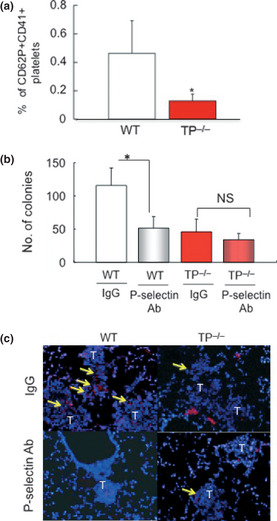
P‐selectin activation through TP signaling enhances pulmonary metastasis. (a) The percentage of P‐selectin+/CD41+ platelets in peripheral blood 7 days after the injection of B16F1 cells. Data are means ± SD for the number of mice (n = 8). *P < 0.05 vs wild‐type mice (WT; Student's t‐test). (b) A neutralizing P‐selectin monoclonal antibody reduced colony formation in WT, but not thromboxane prostanoid receptor knockout mice (TP −/−). Data are means ± SD for the number of mice (n = 5). *P < 0.05 (Student's t‐test). NS, not significant. (c) Immunohistochemical detection of αIIb in metastatic areas. Treatment of WT with a P‐selectin antibody reduced the attachment of platelets to metastatic tumor cells compared with IgG‐treated WT. Platelet attachment to metastatic tumor cells was attenuated in TP −/−. Treatment of TP −/− with a P‐selectin antibody failed to further inhibit the attachment of platelets. Yellow arrows show platelets. Bars, 50 μm.
