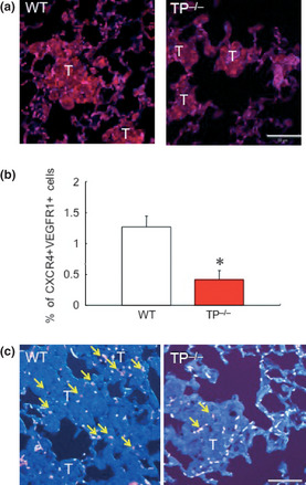
Figure 6.

Thromboxane prostanoid receptor (TP) signaling induces the mobilization of VEGFR1+ CXCR4+ cells in the peripheral blood and homing into lung tissues during the colonization of B16F1 cells. (a) Immunohistochemical detection of vascular endothelial growth factor (VEGF) and CD31 in metastatic areas. Wild‐type (WT) mice express VEGF (red) and CD31 (blue) in metastasis area compared with TP knockout mice (TP −/−) mice 7 days after B16F1 cell injection. Bars, 50 μm. (b) The percentage of VEGFR1+ CXCR4+ cells in the peripheral blood 7 days after B16F1 cell injection. Data are means ± SD for the number of mice (n = 8). *P < 0.05 vs WT (Student's t‐test). (c) Reduced homing of CXCR4+ VEGFR1+ cells to the lung tissue in TP −/− 7 days after the injection of B16F1 cells. Yellow arrows show CXCR4 and VEGFR1 double‐positive cells (magenta). T indicates metastatic area. All images are representative of three independent samples. Bars, 50 μm.
