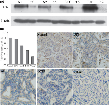
Figure 1.

Expression of the testin (TES) gene in breast cancer tissues. (A) TES protein levels in four different pairs of tumor tissues (T) and their matched non‐tumor tissues (N) analyzed by Western blot. (B) Expression of TES by immunohistochemistry in breast pre‐cancerous lesions and cancer; TES staining was mainly localized in the cytoplasm of breast cells. ADH, atypical ductal hyperplasia; DCIS, ductal carcinoma in situ; UDH, usual ductal hyperplasia without atypia.
