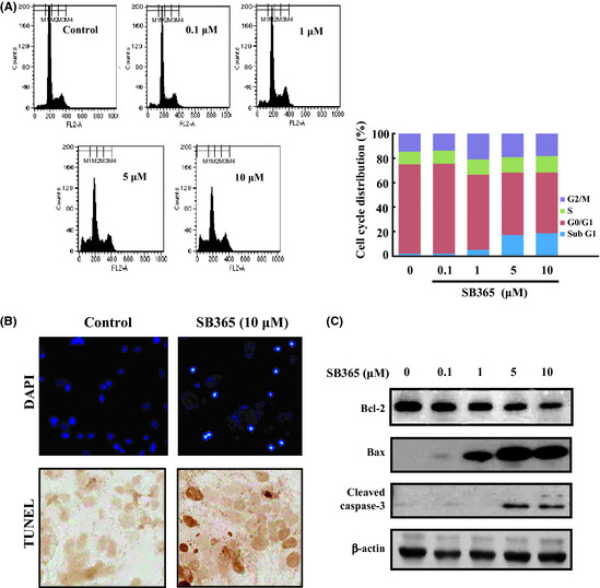
Figure 2.

Effect of SB365 on apoptosis of Huh‐7 cells. (A) Huh‐7 cells were treated with SB365 (0, 0.1, 1, 5 and 10 μM) for 24 h, stained with propidium iodide (PI) and analyzed on a FACSCalibur flow cytometer. (B) The induction of apoptosis by SB365 was conducted by DAPI and TUNEL staining, which were photographed at ×100 and ×400 magnification. (C) The expression of Bcl‐2, Bax and cleaved caspase‐3 were determined by western blotting in cells treated with SB365 at the indicated doses for 24 h.
