
Figure 8.
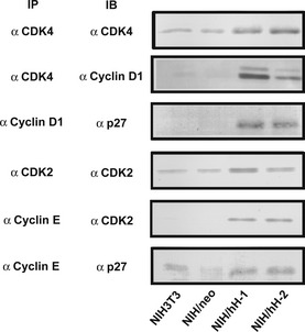
Immunoprecipitation and immunoblot analysis of cyclin D1‐CDK4 and cyclin E‐CDK2 complexes. Equal amounts of lysate proteins from NIH3T3, NIH/neo and two CD98hc‐transfected clones cultured in 0.2% FBS‐medium were treated with indicated antibodies (IP) for immunoprecipitation. Immunoprecipitates were subjected to SDS‐PAGE, blotted onto membranes and stained with indicated antibodies.